Eye
Anatomy of the Eye
The human eye is a complex organ that allows for the perception of visual stimuli. It is composed of several different structures, each with a specific role in the process of vision.


The Eyeball
The eyeball is the spherical structure that houses the main components of the visual system. It is divided into two segments: the anterior segment, which includes the cornea and the lens, and the posterior segment, which contains the vitreous humor, the retina, and the optic nerve.
The Cornea
The cornea is the transparent front part of the eye that covers the iris, pupil, and anterior chamber. It refracts, or bends, light that enters the eye, contributing to the eye's overall optical power.
The Iris and Pupil
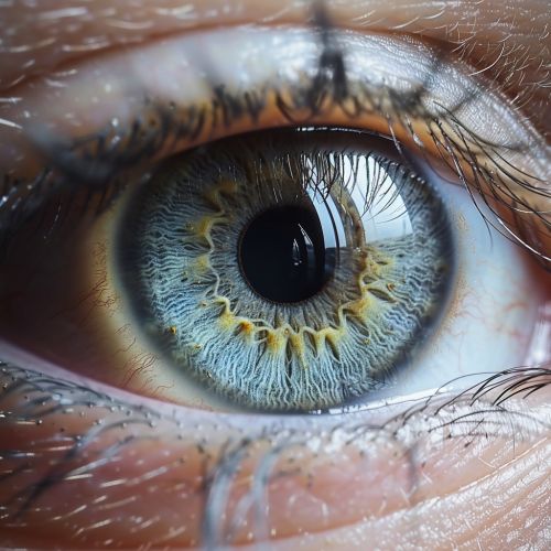
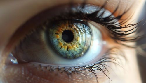
The iris is the colored part of the eye, and it controls the size of the pupil, the opening that allows light to enter the eye. The iris adjusts the size of the pupil in response to changes in light intensity.
The Lens
The lens is a transparent, biconvex structure located behind the iris. It further refracts incoming light to focus it onto the retina.
The Retina
The retina is the innermost layer of the eye. It contains photoreceptor cells that convert light into electrical signals. These signals are then sent to the brain via the optic nerve, resulting in visual perception.
Physiology of Vision
The process of vision begins when light enters the eye through the cornea. The light is then refracted by the cornea and the lens, focusing it onto the retina. The photoreceptor cells in the retina convert the light into electrical signals, which are then transmitted to the brain via the optic nerve. The brain interprets these signals as visual images.
Photoreception
Photoreception is the process by which the eye detects light. This process is carried out by two types of photoreceptor cells in the retina: rods and cones. Rods are responsible for vision in low light conditions, while cones are responsible for color vision and detail.
Visual Processing
Once the light has been converted into electrical signals by the photoreceptor cells, these signals are processed by other cells in the retina. The processed signals are then sent to the brain via the optic nerve, where they are interpreted as visual images.
Eye Health and Disorders
The eye is susceptible to a variety of disorders and conditions that can impair vision. These include refractive errors, such as myopia (nearsightedness) and hyperopia (farsightedness), as well as diseases like glaucoma and macular degeneration.
Refractive Errors
Refractive errors occur when the eye's shape prevents light from focusing directly on the retina. The most common types of refractive errors are myopia, hyperopia, astigmatism, and presbyopia.
Eye Diseases
There are many diseases that can affect the eye and impair vision. These include glaucoma, a condition characterized by damage to the optic nerve, and macular degeneration, a disease that affects the central part of the retina and impairs central vision.
