Extracellular Vesicles
Introduction
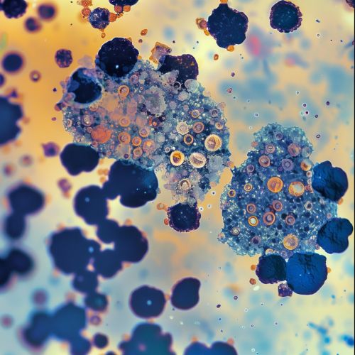
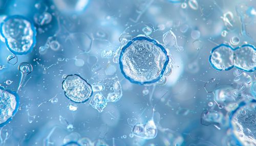
Extracellular vesicles (EVs) are membrane-bound particles released from cells into the extracellular environment. These vesicles play a crucial role in intercellular communication and are involved in various physiological and pathological processes. EVs are heterogeneous in nature and can be classified into different subtypes based on their size, biogenesis, and function. The primary subtypes include exosomes, microvesicles, and apoptotic bodies.
Classification of Extracellular Vesicles
Exosomes
Exosomes are small EVs ranging from 30 to 150 nanometers in diameter. They originate from the endosomal pathway and are released into the extracellular space when multivesicular bodies (MVBs) fuse with the plasma membrane. Exosomes carry a diverse array of biomolecules, including proteins, lipids, and nucleic acids, which can influence recipient cells.
Microvesicles
Microvesicles, also known as ectosomes, are larger than exosomes, typically ranging from 100 to 1000 nanometers. They are formed by the outward budding and fission of the plasma membrane. Microvesicles contain cytoplasmic components and membrane proteins from the parent cell, and their composition reflects the state of the cell at the time of release.
Apoptotic Bodies
Apoptotic bodies are the largest type of EVs, ranging from 500 to 2000 nanometers. They are released during apoptosis, a form of programmed cell death. Apoptotic bodies contain cellular organelles, nuclear fragments, and other cellular debris, and they play a role in the clearance of dying cells by phagocytes.
Biogenesis of Extracellular Vesicles
The biogenesis of EVs involves complex cellular processes that vary depending on the type of vesicle. Exosome formation begins with the inward budding of the endosomal membrane to form intraluminal vesicles (ILVs) within MVBs. These MVBs can either fuse with lysosomes for degradation or with the plasma membrane to release ILVs as exosomes. Microvesicles are generated through the direct outward budding of the plasma membrane, a process regulated by cytoskeletal rearrangements and lipid composition. Apoptotic bodies are produced during the late stages of apoptosis when the cell undergoes fragmentation.
Composition of Extracellular Vesicles
EVs are composed of a lipid bilayer membrane that encapsulates various biomolecules. The lipid composition of EVs includes phospholipids, cholesterol, and sphingolipids, which contribute to their stability and functionality. Proteins found in EVs include tetraspanins (e.g., CD9, CD63, CD81), heat shock proteins, and enzymes. EVs also carry nucleic acids, such as mRNA, microRNA, and DNA, which can be transferred to recipient cells and modulate gene expression.
Functions of Extracellular Vesicles
EVs are involved in numerous biological processes, including immune response, tissue repair, and cancer progression. They facilitate intercellular communication by transferring bioactive molecules between cells. In the immune system, EVs can present antigens to immune cells, modulate immune responses, and contribute to the spread of viral infections. In tissue repair, EVs derived from stem cells promote regeneration and healing by delivering growth factors and other regenerative molecules. In cancer, EVs can influence tumor progression by promoting angiogenesis, metastasis, and immune evasion.


Clinical Applications of Extracellular Vesicles
EVs hold significant potential for clinical applications in diagnostics and therapeutics. Their ability to carry and protect biomolecules makes them ideal candidates for biomarkers in liquid biopsies. EVs can be isolated from various body fluids, such as blood, urine, and cerebrospinal fluid, and analyzed for disease-specific signatures. In therapeutics, EVs can be engineered to deliver drugs, nucleic acids, or proteins to target cells, offering a novel approach for treating diseases such as cancer, neurodegenerative disorders, and cardiovascular diseases.
Isolation and Characterization of Extracellular Vesicles
The isolation of EVs from biological samples involves several techniques, including differential ultracentrifugation, size-exclusion chromatography, and immunoaffinity capture. Each method has its advantages and limitations in terms of yield, purity, and scalability. Characterization of EVs typically involves assessing their size, concentration, and molecular composition using techniques such as nanoparticle tracking analysis (NTA), dynamic light scattering (DLS), and flow cytometry. Proteomic and genomic analyses are also employed to identify the cargo carried by EVs.
Challenges and Future Directions
Despite the promising potential of EVs, several challenges remain in their study and application. Standardization of isolation and characterization methods is crucial for reproducibility and comparability of results. Understanding the mechanisms underlying EV biogenesis, cargo selection, and uptake by recipient cells is essential for harnessing their full therapeutic potential. Future research should focus on elucidating the role of EVs in various diseases, developing scalable production methods, and exploring their use in personalized medicine.
See Also
- Endosomal Pathway
- Apoptosis
- Intercellular Communication
- Liquid Biopsy
- Nanoparticle Tracking Analysis
- Stem Cell Therapy
