Electrocardiography
Introduction
Electrocardiography is a commonly used, non-invasive procedure for recording electrical changes in the heart. The term is derived from the Greek words 'electro' referring to its relation to electrical activity, 'cardio' meaning heart, and 'graph' meaning to write. Any disturbance in the normal electrical activity of the heart manifests as changes in the ECG.
History
The history of electrocardiography can be traced back to the 19th century. An Italian physician named Luigi Galvani was the first to discover that the heart produces electrical activity. However, it was not until the late 19th and early 20th centuries that Dutch physiologist Willem Einthoven developed the first practical ECG machine, a development for which he was awarded the Nobel Prize in Medicine in 1924.
Basic Principles
The basic principle of electrocardiography involves the recording of electrical activity of the heart over a period of time as detected by electrodes placed on the skin. These electrodes detect the small electrical changes that are a consequence of cardiac muscle depolarization followed by repolarization during each cardiac cycle (heartbeat).
Electrocardiogram
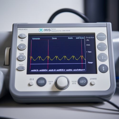
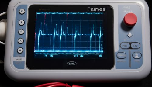
An electrocardiogram (ECG) is the graphical record produced by an electrocardiograph, which records the electrical activity of the heart over time. Its name comes from the Greek words 'elektor' meaning 'amber', which was the source of the word 'electricity', 'kardia' meaning 'heart', and 'gramma' meaning 'something written'.
Components of ECG
An ECG is composed of waves, intervals, and complexes which represent specific events during the heart's electrical cycle. The P wave represents atrial depolarization, the QRS complex represents ventricular depolarization, and the T wave represents ventricular repolarization.
Interpretation of ECG
Interpretation of the ECG is fundamentally about understanding the electrical conduction system of the heart. Normal cardiac rhythm originates in the sinoatrial (SA) node and follows a set path through the atria, atrioventricular (AV) node, bundle of His, bundle branches, and Purkinje fibers. Disturbances or abnormalities in this conduction pathway can lead to characteristic changes in the ECG.
Clinical Significance
Electrocardiography is a vital tool in medical practice, used in the evaluation of patients with suspected heart disease. It can provide information on the heart rate, rhythm, and morphology of the electrical activity, which can aid in the diagnosis of various cardiac conditions such as arrhythmias, myocardial infarction, and more.
Limitations and Challenges
While electrocardiography is a valuable tool, it is not without limitations. The ECG provides a two-dimensional representation of a three-dimensional phenomenon, which can sometimes lead to misinterpretation. Moreover, certain conditions may not produce changes in the ECG until they are quite advanced.
Future Directions
The future of electrocardiography lies in technological advancements and their application in clinical practice. Innovations in wearable technology, machine learning, and artificial intelligence are poised to revolutionize the field of electrocardiography.
See Also


