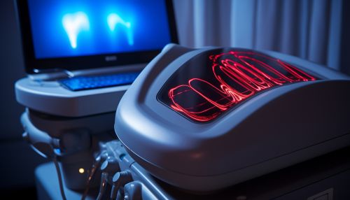
Echocardiography
Overview
Echocardiography is a diagnostic test that uses ultrasound waves to produce images of the heart. This non-invasive procedure allows doctors to monitor the functioning of the heart and its valves, making it a crucial tool in the diagnosis of heart disease. The images generated by an echocardiogram provide valuable information about the size, shape, and movement of the heart and its components, such as the heart valves, the septum (the wall separating the left and right heart chambers), and the walls of the heart chambers.


History
The development of echocardiography has been a significant milestone in the field of cardiology. The first practical application of ultrasound in medical diagnosis was in the 1940s, and by the 1950s, ultrasound was being used for clinical purposes. However, it was not until the 1960s that the first echocardiograms were produced. The technique was initially limited to the detection of mitral stenosis, a condition in which the heart's mitral valve narrows, restricting blood flow.
Types of Echocardiography
There are several types of echocardiography, each with its own specific uses and advantages. These include Transthoracic echocardiography, Transesophageal echocardiography, and Stress echocardiography.
Transthoracic Echocardiography
Transthoracic echocardiography (TTE) is the most common type of echocardiography. It involves the placement of an ultrasound transducer on the chest wall of the patient to obtain images of the heart. TTE is non-invasive, painless, and provides a wealth of information about the heart's structure and function.
Transesophageal Echocardiography
Transesophageal echocardiography (TEE) involves the insertion of a specialized probe with an ultrasound transducer at its tip into the patient's esophagus. This allows for a closer and more accurate image of the heart, particularly the back structures, such as the left atrium, which are not as easily visualized with TTE.
Stress Echocardiography
Stress echocardiography is used to assess the heart's response to stress or exercise. The patient exercises on a treadmill or stationary bicycle while the echocardiogram is performed. Alternatively, medication may be used to stimulate the heart in a way similar to exercise.
Procedure
The procedure for an echocardiogram varies depending on the type of echocardiography being performed. However, in general, the patient will be asked to remove any clothing from the waist up and to lie down on an examination table. Electrodes will be attached to the patient's chest to monitor the heart's activity.
For a TTE, a transducer (a small device that produces sound waves) is moved around on the chest. The transducer sends sound waves into the body, which bounce off the heart and return to the transducer. The sound waves are then converted into images on a screen.
In a TEE, the transducer is attached to a thin tube that is passed down the patient's throat and into the esophagus. The images obtained provide a detailed picture of the heart's structure and function.
During a stress echocardiogram, the patient will either exercise or be given medication to make the heart beat faster and harder. An echocardiogram is then performed both before and after the heart is stressed.
Applications
Echocardiography has a wide range of applications and is used to diagnose and monitor a variety of heart conditions. These include coronary artery disease, heart failure, congenital heart disease, and problems with the heart valves. Echocardiography can also be used to detect blood clots or tumors within the heart, to measure the size and shape of the heart, to assess how well the heart pumps blood, and to evaluate the function of the heart valves.
Limitations and Risks
While echocardiography is a valuable diagnostic tool, it does have some limitations. For instance, the quality of the images obtained can be affected by a number of factors, including the patient's body size, the presence of lung disease, and the patient's ability to hold still during the procedure. Additionally, while echocardiography is generally considered safe, there are some risks associated with certain types of echocardiography. For example, TEE is an invasive procedure and carries risks such as throat discomfort, bleeding, and, in rare cases, perforation of the esophagus.
Future Developments
The field of echocardiography continues to evolve, with new technologies and techniques being developed to improve the quality of images and the information they provide. For example, three-dimensional echocardiography is a relatively new technique that allows for the acquisition of three-dimensional images of the heart, providing even more detailed information about its structure and function.
