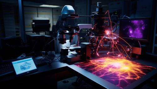
Confocal Microscopy
Introduction
Confocal microscopy is a technique in optical microscopy that uses point illumination and a pinhole in an optically conjugate plane in front of the detector to eliminate out-of-focus signal. The primary advantage of this technique is that it allows imaging of thick specimens with nearly the same resolution as with thin specimens.


History
The principle of confocal imaging was patented by Marvin Minsky in 1957. At the time, Minsky was seeking to improve the resolution and contrast of images captured by light microscopes, which were limited by the large amount of out-of-focus light. His patent included the key elements of confocal microscopy, such as the use of point illumination and a pinhole to eliminate out-of-focus light.
Principle
Confocal microscopy involves the use of point illumination and a pinhole in an optically conjugate plane in front of the detector to eliminate out-of-focus light. This is achieved by using a laser to illuminate a single point in the specimen, and then scanning the laser across the specimen to image the entire area. The light from the illuminated point is then collected by a detector, and the intensity of the detected light is used to create an image.
Applications
Confocal microscopy has a wide range of applications in various fields, including biology, medicine, and materials science. In biology, it is used to study cellular structures and dynamics, while in medicine, it is used for imaging tissues and cells in vivo. In materials science, it is used to characterize the surface topology of materials.
Advantages and Limitations
The main advantage of confocal microscopy is its ability to produce high-resolution images of thick specimens. This is because the use of a pinhole to eliminate out-of-focus light allows for the imaging of a thin slice of the specimen, known as an optical section. This optical sectioning capability makes confocal microscopy ideal for three-dimensional imaging of thick specimens.
However, confocal microscopy also has several limitations. One of the main limitations is the relatively slow imaging speed, which is due to the need to scan the laser across the specimen. Additionally, the use of a laser can cause photobleaching and phototoxicity, which can damage the specimen.
Techniques
There are several techniques in confocal microscopy, including laser scanning confocal microscopy, spinning disk confocal microscopy, and two-photon excitation microscopy. Each of these techniques has its own advantages and limitations, and the choice of technique depends on the specific requirements of the application.
Future Directions
The field of confocal microscopy continues to evolve, with ongoing research aimed at improving the performance and capabilities of confocal microscopes. Future directions in this field include the development of new imaging techniques, the integration of confocal microscopy with other imaging modalities, and the application of artificial intelligence for image analysis.
