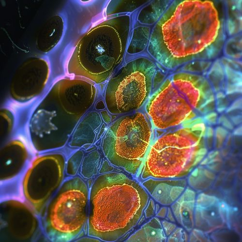
Cone cell
Introduction
Cone cells, or cones, are one of the two types of photoreceptor cells that are in the retina of the eye which are responsible for color vision. They are also able to function in less intense light than the other type of visual photoreceptor, rod cells. Cones are concentrated in the fovea centralis, the center of the macula, where visual acuity (sharpness) is the highest. They are less sensitive to light than the rod cells in the retina (which support vision at low light levels), but they allow the perception of color and are responsible for high spatial acuity.


Structure
Cone cells are somewhat shorter than rods, but wider and tapered, and are much less numerous than rods in most parts of the retina, but greatly outnumber rods in the fovea. Structurally, cone cells have a cone-like shape at one end where a pigment filters incoming light, giving them their different response curves. They are typically 40-50 μm long, and their diameter varies from 0.5 to 4.0 μm, being smallest and most tightly packed at the center of the eye at the fovea.
Function
Cone cells are adapted for color vision, photopic vision (vision under well-lit conditions), and are responsible for high spatial acuity. The S (for short-wavelength) cones are most sensitive to light that we perceive as the color blue. The M (for medium-wavelength) cones are most sensitive to light that we perceive as the color green. The L (for long-wavelength) cones are most sensitive to light that we perceive as the color red.
Types of Cone Cells
There are three types of cones that differ in the wavelengths of light to which they respond. They are called L-cones, M-cones and S-cones. Each type of cone is named for the portion of the spectrum of light to which it is sensitive: short, medium, or long wavelength.
L-cones
The L-cones, or long-wavelength cones, or red cones, are most sensitive to light that is perceived as greenish-yellow, although it is often described as red, and are insensitive to short-wavelength light, the so-called "blue" light.
M-cones
The M-cones, or medium-wavelength cones, or green cones, are most sensitive to light that is perceived as green, but is also sensitive to other wavelengths.
S-cones
The S-cones, or short-wavelength cones, or blue cones, are most sensitive to light that is perceived as blue and are insensitive to long-wavelength light, the so-called "red" light.
Distribution
The human retina contains about 6 million cone cells. In the fovea, which is the area of clearest vision, the layers of the retina spread aside to let light fall directly on the cones. These cells are most densely packed in this tiny pit in the center of the retina that provides the clearest, most focused vision.
Cone Cells and Color Vision
The ability to see different colors is due to the variable ratio of the three types of cones stimulated by different colors of light. The brain combines the information from each type of receptor to give rise to different perceptions of different wavelengths of light.
Diseases and Disorders
There are several hereditary diseases that can affect the cone cells and disrupt color vision. The most common is color blindness. Other disorders like achromatopsia (total color blindness) can also occur. In addition, age-related macular degeneration, a disease that results in a loss of central vision, is caused by damage to the cone cells in the macula.
