Centriole
Overview
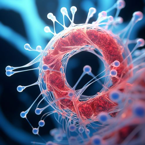
A Centriole is a cylindrical cellular structure composed primarily of a protein called tubulin. It is found in most eukaryotic cells, with the notable exception of higher plants. Centrioles are typically found in pairs, arranged at right angles to each other, and are located near the nucleus in a region called the centrosome. They play a crucial role in cell division, acting as the primary microtubule organizing center (MTOC) of the cell.


Structure
The structure of a centriole is remarkably consistent across different cell types and species. Each centriole is a barrel-shaped structure approximately 500 nanometers in length and 200 nanometers in diameter. The wall of the centriole is composed of nine triplet microtubules, each of which is made up of three individual tubulin proteins arranged in a specific pattern. This arrangement of microtubules is often referred to as a "9+0" pattern, indicating the nine triplet microtubules and the absence of a central pair of microtubules.
Function
The primary function of the centriole is to organize the microtubules of the cell during cell division. During this process, the centrioles replicate and move to opposite ends of the cell, forming the poles of the mitotic spindle. The microtubules, which are essential for the separation of chromosomes, then radiate from the centrioles.
In addition to their role in cell division, centrioles are also involved in the formation of cilia and flagella. These are long, hair-like structures that protrude from the cell surface and are involved in cell movement and the transport of materials across the cell surface. The centrioles form the basal bodies, which are the structures at the base of cilia and flagella from which the microtubules extend.
Biogenesis and Replication
Centriole biogenesis, the process by which new centrioles are formed, is a tightly regulated process that occurs in coordination with the cell cycle. During the S phase of the cell cycle, each centriole begins to duplicate by forming a procentriole, a small bud that grows out from the side of the parent centriole. The procentriole elongates and matures during the G2 phase and mitosis, eventually becoming a fully formed centriole by the end of the cell cycle.
The exact mechanisms by which centrioles replicate are still not fully understood, but it is known that several proteins, including PLK4, SAS-6, and STIL, play crucial roles in this process. Disruptions in centriole replication can lead to a variety of cellular defects, including abnormal cell division and ciliopathies, a group of diseases caused by defects in cilia formation or function.
Clinical Significance
Given their essential role in cell division, centrioles have been implicated in a number of diseases, most notably cancer. Abnormalities in centriole number or structure can lead to errors in chromosome segregation during cell division, resulting in aneuploidy, a condition in which cells have an abnormal number of chromosomes. Aneuploidy is a hallmark of many types of cancer, suggesting that defects in centriole function may contribute to tumorigenesis.
In addition to cancer, defects in centriole function have also been implicated in a number of genetic disorders known as ciliopathies. These include diseases such as polycystic kidney disease, Bardet-Biedl syndrome, and primary ciliary dyskinesia. In these conditions, defects in centriole function lead to abnormalities in cilia formation or function, resulting in a wide range of symptoms including kidney disease, obesity, and respiratory problems.
