Cardiomyocytes
Introduction
Cardiomyocytes, also known as cardiac muscle cells, are the primary cell type found in the heart. These cells are responsible for the contractile function of the heart, which enables it to pump blood throughout the body. Cardiomyocytes are unique in their structure and function, differing significantly from other types of muscle cells in the body.


Structure
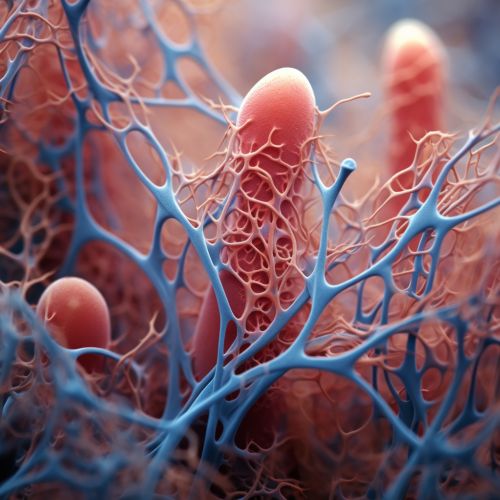
The structure of cardiomyocytes is complex and highly specialized to facilitate their function. They are elongated, cylindrical cells that are approximately 100-150 micrometers in length and 10-30 micrometers in diameter. Unlike other muscle cells, cardiomyocytes contain a single, centrally located nucleus.
The cytoplasm of cardiomyocytes, known as the sarcoplasm, contains numerous mitochondria to meet the high energy demands of these cells. The sarcoplasm is also filled with myofibrils, which are bundles of protein filaments that are responsible for the contractile properties of the cell.
The surface of cardiomyocytes is covered with a specialized cell membrane known as the sarcolemma. The sarcolemma has deep invaginations called T-tubules that allow for the rapid spread of electrical signals throughout the cell.
Function
The primary function of cardiomyocytes is to contract and relax, thereby enabling the heart to pump blood. This contractile activity is regulated by a complex sequence of electrical and chemical events known as the cardiac action potential.
The cardiac action potential begins with the rapid influx of sodium ions into the cell, causing it to depolarize. This is followed by a slower influx of calcium ions, which triggers the release of more calcium from the sarcoplasmic reticulum, a specialized organelle within the cell. The increased concentration of calcium ions within the cell activates the myofibrils, causing them to contract.
Following contraction, the cell repolarizes as potassium ions exit the cell and calcium ions are pumped back into the sarcoplasmic reticulum. This allows the myofibrils to relax, preparing the cell for the next contraction.
Regulation
The contractile activity of cardiomyocytes is tightly regulated by various factors. These include the autonomic nervous system, hormones, and local factors within the heart itself.
The autonomic nervous system exerts a major influence on the heart through the sympathetic and parasympathetic branches. The sympathetic nervous system increases heart rate and contractility, while the parasympathetic nervous system decreases these parameters.
Hormones such as adrenaline and noradrenaline, released by the adrenal glands, also increase heart rate and contractility. On the other hand, hormones like acetylcholine and atrial natriuretic peptide decrease these parameters.
Local factors within the heart, such as changes in blood volume and pressure, can also influence the activity of cardiomyocytes. For example, an increase in blood volume within the heart can stretch the cardiomyocytes, leading to an increase in their contractile force. This is known as the Frank-Starling law of the heart.
Pathology
Cardiomyocytes are susceptible to various diseases and conditions. For example, myocardial infarction, commonly known as a heart attack, occurs when the blood supply to a part of the heart is blocked, leading to the death of cardiomyocytes in that area. This can result in permanent damage to the heart and can lead to heart failure.
Cardiomyocytes can also be affected by genetic conditions such as hypertrophic cardiomyopathy, where the cells become abnormally large, leading to thickening of the heart walls. This can impair the heart's ability to pump blood effectively.
Other conditions that can affect cardiomyocytes include viral infections, autoimmune diseases, and certain drugs and toxins. In many of these cases, damage to the cardiomyocytes can lead to arrhythmias, or irregular heart rhythms.
Research and Future Directions
Research into cardiomyocytes is ongoing and holds promise for the development of new treatments for heart disease. One area of research is the study of cardiomyocyte regeneration. Unlike many other types of cells in the body, cardiomyocytes have a limited ability to divide and regenerate. However, recent studies have suggested that it may be possible to stimulate cardiomyocyte regeneration, which could potentially be used to repair damaged heart tissue.
Another area of research is the use of cardiomyocytes in drug testing. By growing cardiomyocytes in the lab, researchers can test the effects of new drugs on these cells before they are tested in humans. This can help to predict potential side effects and improve the safety of new treatments.
