Choroiditis: Difference between revisions
(Created page with "== Overview == Choroiditis is an inflammatory condition affecting the choroid, a vascular layer of the eye situated between the retina and the sclera. This condition can lead to significant visual impairment if not diagnosed and treated promptly. Choroiditis can be classified into various subtypes based on the etiology, clinical presentation, and histopathological findings. Understanding the underlying causes, clinical manifestations, diagnostic methods, and treatment o...") |
No edit summary |
||
| (One intermediate revision by the same user not shown) | |||
| Line 25: | Line 25: | ||
=== Imaging Studies === | === Imaging Studies === | ||
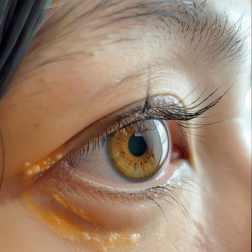
[[Image:Detail-79569.jpg|thumb|center|Fundus photograph showing choroiditis lesions in the eye.|class=only_on_mobile]] | |||
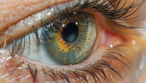
[[Image:Detail-79570.jpg|thumb|center|Fundus photograph showing choroiditis lesions in the eye.|class=only_on_desktop]] | |||
Imaging modalities such as fundus photography, fluorescein angiography, and optical coherence tomography (OCT) are invaluable in diagnosing choroiditis. Fundus photography helps document the appearance of choroidal lesions, while fluorescein angiography can reveal areas of leakage and ischemia. OCT provides detailed cross-sectional images of the retina and choroid, aiding in the assessment of structural changes. | Imaging modalities such as fundus photography, fluorescein angiography, and optical coherence tomography (OCT) are invaluable in diagnosing choroiditis. Fundus photography helps document the appearance of choroidal lesions, while fluorescein angiography can reveal areas of leakage and ischemia. OCT provides detailed cross-sectional images of the retina and choroid, aiding in the assessment of structural changes. | ||
Latest revision as of 00:13, 19 May 2024
Overview
Choroiditis is an inflammatory condition affecting the choroid, a vascular layer of the eye situated between the retina and the sclera. This condition can lead to significant visual impairment if not diagnosed and treated promptly. Choroiditis can be classified into various subtypes based on the etiology, clinical presentation, and histopathological findings. Understanding the underlying causes, clinical manifestations, diagnostic methods, and treatment options is crucial for effective management.
Anatomy of the Choroid
The choroid is a critical component of the uveal tract, which also includes the iris and ciliary body. It is composed of blood vessels, connective tissue, and melanocytes. The primary function of the choroid is to provide oxygen and nutrients to the outer layers of the retina. The choroid is divided into several layers: the suprachoroid, large vessel layer (Haller's layer), medium vessel layer (Sattler's layer), and the choriocapillaris. Each layer plays a distinct role in maintaining ocular health and function.
Etiology
Choroiditis can be caused by a variety of factors, including infectious agents, autoimmune disorders, and idiopathic conditions. The most common infectious causes include Toxoplasmosis, Tuberculosis, and Syphilis. Autoimmune conditions such as Vogt-Koyanagi-Harada disease, Sarcoidosis, and Behçet's disease can also lead to choroiditis. Idiopathic choroiditis, where no specific cause is identified, is another significant category.
Clinical Manifestations
The clinical presentation of choroiditis can vary widely depending on the underlying cause and extent of inflammation. Common symptoms include blurred vision, floaters, photophobia, and visual field defects. In severe cases, patients may experience pain and redness in the affected eye. Fundoscopic examination often reveals yellow-white lesions in the choroid, retinal detachment, and vitreous inflammation.
Diagnosis
Diagnosing choroiditis involves a combination of clinical evaluation, imaging studies, and laboratory tests.
Clinical Evaluation
A thorough history and physical examination are essential. Ophthalmologists often use slit-lamp biomicroscopy and indirect ophthalmoscopy to assess the extent of inflammation and identify characteristic lesions.
Imaging Studies


Imaging modalities such as fundus photography, fluorescein angiography, and optical coherence tomography (OCT) are invaluable in diagnosing choroiditis. Fundus photography helps document the appearance of choroidal lesions, while fluorescein angiography can reveal areas of leakage and ischemia. OCT provides detailed cross-sectional images of the retina and choroid, aiding in the assessment of structural changes.
Laboratory Tests
Laboratory investigations are tailored based on the suspected etiology. Serological tests for infectious agents, autoimmune markers, and chest X-rays or CT scans for systemic diseases like tuberculosis and sarcoidosis may be required.
Treatment
The treatment of choroiditis depends on the underlying cause and severity of the condition.
Infectious Choroiditis
For infectious choroiditis, antimicrobial therapy is the mainstay of treatment. For example, Toxoplasmosis choroiditis is treated with a combination of pyrimethamine, sulfadiazine, and folinic acid. Tuberculous choroiditis requires anti-tubercular therapy, while syphilitic choroiditis is treated with penicillin.
Autoimmune Choroiditis
Autoimmune choroiditis often necessitates immunosuppressive therapy. Corticosteroids are commonly used to reduce inflammation. In refractory cases, immunomodulatory agents such as methotrexate, azathioprine, or biologics like infliximab may be employed.
Idiopathic Choroiditis
Idiopathic choroiditis is managed based on the severity of symptoms and the extent of inflammation. Corticosteroids are typically the first line of treatment, with immunosuppressive agents reserved for more severe or recurrent cases.
Prognosis
The prognosis of choroiditis varies depending on the cause and promptness of treatment. Infectious choroiditis generally has a good prognosis if treated early. Autoimmune and idiopathic forms can be more challenging to manage and may require long-term therapy to prevent recurrences and complications.
Complications
Potential complications of choroiditis include Cataract, Glaucoma, and Retinal detachment. Chronic inflammation can lead to scarring and permanent vision loss. Regular follow-up and monitoring are essential to detect and manage these complications promptly.
Conclusion
Choroiditis is a complex condition with diverse etiologies and clinical presentations. Early diagnosis and appropriate treatment are crucial to prevent vision loss and improve outcomes. Ongoing research and advances in imaging and therapeutic modalities continue to enhance our understanding and management of this challenging condition.
