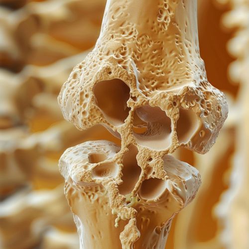
Epiphyseal plate
Epiphyseal Plate
The epiphyseal plate, also known as the growth plate or physis, is a hyaline cartilage plate located in the metaphysis at each end of a long bone. This structure is crucial for the longitudinal growth of bones during childhood and adolescence. The epiphyseal plate is present in children and adolescents; in adults, who have stopped growing, the plate is replaced by an epiphyseal line.


Structure
The epiphyseal plate is composed of four distinct zones: the resting zone, the proliferative zone, the hypertrophic zone, and the calcification zone. Each of these zones plays a specific role in the process of endochondral ossification, which is the method by which bones grow in length.
- **Resting Zone**: This zone is closest to the epiphysis and contains small, scattered chondrocytes. These cells are relatively inactive but serve as a reserve of cells that can be recruited for proliferation.
- **Proliferative Zone**: In this zone, chondrocytes undergo rapid mitosis, leading to an increase in the number of cells. These cells are arranged in columns parallel to the long axis of the bone.
- **Hypertrophic Zone**: Chondrocytes in this zone cease to divide and begin to enlarge. The lacunae surrounding these cells also expand, creating a larger matrix.
- **Calcification Zone**: This zone is characterized by the calcification of the cartilage matrix. Chondrocytes die and are replaced by osteoblasts, which lay down bone matrix.
Function
The primary function of the epiphyseal plate is to facilitate the longitudinal growth of bones. This is achieved through the process of endochondral ossification, where cartilage is progressively replaced by bone tissue. Growth hormone and other factors such as thyroid hormone and sex hormones regulate this process.
Clinical Significance
The epiphyseal plate is a critical structure in pediatric orthopedics. Injuries to the growth plate can result in growth disturbances, leading to limb length discrepancies or angular deformities. Conditions such as achondroplasia and gigantism are associated with abnormalities in the growth plate.
- **Growth Plate Fractures**: These are classified using the Salter-Harris system, which ranges from Type I to Type V, based on the severity and location of the fracture.
- **Growth Plate Disorders**: Conditions such as osteochondritis dissecans and slipped capital femoral epiphysis involve the epiphyseal plate and can significantly impact bone growth and development.
Hormonal Regulation
The activity of the epiphyseal plate is regulated by a complex interplay of hormones. Growth hormone, produced by the anterior pituitary gland, stimulates the proliferation of chondrocytes in the proliferative zone. Thyroid hormones are essential for the proper maturation of chondrocytes, while sex hormones such as estrogen and testosterone accelerate the closure of the growth plate during puberty.
- **Growth Hormone**: Deficiency or excess of growth hormone can lead to conditions such as dwarfism or acromegaly, respectively.
- **Thyroid Hormones**: Hypothyroidism can result in delayed bone growth and development, while hyperthyroidism can lead to accelerated growth plate closure.
- **Sex Hormones**: Estrogen has a more potent effect on the closure of the growth plate compared to testosterone, which is why females typically stop growing earlier than males.
Molecular Mechanisms
At the molecular level, the regulation of the epiphyseal plate involves various signaling pathways and transcription factors. The Indian hedgehog (Ihh) and parathyroid hormone-related protein (PTHrP) signaling pathways are crucial for the coordination of chondrocyte proliferation and differentiation.
- **Ihh Signaling**: Indian hedgehog is produced by prehypertrophic and early hypertrophic chondrocytes and regulates the proliferation of chondrocytes in the proliferative zone.
- **PTHrP Signaling**: Parathyroid hormone-related protein maintains chondrocytes in a proliferative state and delays their differentiation into hypertrophic chondrocytes.
- **Transcription Factors**: Sox9, Runx2, and Osterix are key transcription factors involved in chondrocyte differentiation and the transition from cartilage to bone.
Pathophysiology
Disorders of the epiphyseal plate can result from genetic mutations, hormonal imbalances, or physical trauma. These disorders can lead to abnormal bone growth and development, resulting in various clinical conditions.
- **Genetic Disorders**: Mutations in genes such as FGFR3 (fibroblast growth factor receptor 3) can lead to conditions like achondroplasia, characterized by short stature and disproportionately short limbs.
- **Hormonal Imbalances**: Conditions such as hypopituitarism and hyperpituitarism can affect the growth plate's function, leading to growth deficiencies or excesses.
- **Traumatic Injuries**: Physical trauma to the growth plate can result in fractures that disrupt normal bone growth. Early diagnosis and appropriate treatment are essential to minimize long-term complications.
Imaging and Diagnosis
The evaluation of the epiphyseal plate typically involves imaging techniques such as X-rays, MRI, and CT scans. These modalities provide detailed information about the structure and integrity of the growth plate.
- **X-rays**: Standard radiographs are commonly used to assess the growth plate's appearance and detect any fractures or abnormalities.
- **MRI**: Magnetic resonance imaging provides high-resolution images of the growth plate and surrounding soft tissues, making it useful for diagnosing conditions like osteochondritis dissecans.
- **CT Scans**: Computed tomography offers detailed cross-sectional images of the bone and growth plate, aiding in the evaluation of complex fractures and deformities.
Treatment
The treatment of epiphyseal plate injuries and disorders depends on the underlying cause and severity of the condition. Management strategies may include conservative measures, surgical intervention, or hormonal therapy.
- **Conservative Treatment**: Mild growth plate injuries may be managed with immobilization, physical therapy, and close monitoring.
- **Surgical Intervention**: Severe fractures or deformities may require surgical procedures such as open reduction and internal fixation, osteotomy, or epiphysiodesis.
- **Hormonal Therapy**: Conditions resulting from hormonal imbalances may be treated with hormone replacement therapy or medications that modulate hormone levels.
Prognosis
The prognosis for individuals with epiphyseal plate injuries or disorders varies based on the specific condition and the effectiveness of treatment. Early diagnosis and appropriate management are crucial for optimizing outcomes and minimizing long-term complications.
- **Growth Plate Fractures**: With timely and appropriate treatment, most growth plate fractures heal without significant complications. However, severe injuries may result in growth disturbances or deformities.
- **Genetic Disorders**: The prognosis for genetic conditions affecting the growth plate depends on the specific disorder and the availability of targeted therapies. Advances in genetic research and molecular medicine hold promise for improving outcomes in these conditions.
- **Hormonal Imbalances**: Hormonal disorders affecting the growth plate can often be managed effectively with hormone replacement therapy or other medical interventions, leading to improved growth and development.
