Leaf Anatomy: Difference between revisions
No edit summary |
No edit summary |
||
| Line 17: | Line 17: | ||
* **Dichotomous Venation**: Veins fork evenly and progressively from the base of the leaf to the apex, seen in ferns and ginkgo. | * **Dichotomous Venation**: Veins fork evenly and progressively from the base of the leaf to the apex, seen in ferns and ginkgo. | ||
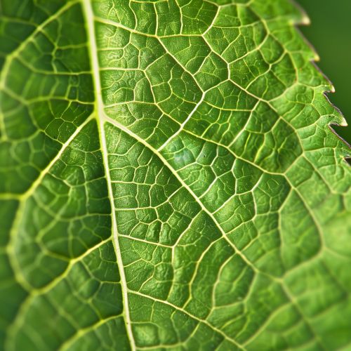
[[Image:Detail-79807.jpg|thumb|center|A close-up image of a green leaf showing its venation pattern.]] | [[Image:Detail-79807.jpg|thumb|center|A close-up image of a green leaf showing its venation pattern.|class=only_on_mobile]] | ||
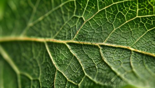
[[Image:Detail-79808.jpg|thumb|center|A close-up image of a green leaf showing its venation pattern.|class=only_on_desktop]] | |||
== Internal Structure == | == Internal Structure == | ||
Latest revision as of 09:58, 20 May 2024
Introduction
Leaf anatomy is a branch of botany that focuses on the structure and organization of leaves. Leaves are the primary photosynthetic organs of most plants, playing a crucial role in converting light energy into chemical energy through the process of photosynthesis. Understanding leaf anatomy is essential for comprehending how plants function and interact with their environment. This article delves into the intricate details of leaf anatomy, exploring various components and their functions.
External Structure
Leaf Morphology
Leaves come in various shapes, sizes, and arrangements, which are collectively referred to as leaf morphology. The primary parts of a leaf include the blade (lamina), petiole, and stipules. The blade is the broad, flat part of the leaf that is primarily responsible for photosynthesis. The petiole is the stalk that attaches the blade to the stem, and stipules are small leaf-like appendages at the base of the petiole.
Leaf Venation
Leaf venation refers to the pattern of veins in a leaf. There are several types of venation, including:
- **Parallel Venation**: Veins run parallel to each other, commonly found in monocots like grasses.
- **Reticulate Venation**: Veins form a network, typical of dicots such as oak and maple trees.
- **Dichotomous Venation**: Veins fork evenly and progressively from the base of the leaf to the apex, seen in ferns and ginkgo.


Internal Structure
Epidermis
The epidermis is the outermost layer of cells covering the leaf. It serves as a protective barrier against physical damage, pathogens, and water loss. The epidermis is usually covered by a waxy cuticle that minimizes water loss. Specialized cells called guard cells are found in the epidermis and regulate the opening and closing of stomata, which are pores that facilitate gas exchange.
Mesophyll
The mesophyll is the inner tissue of the leaf, sandwiched between the upper and lower epidermis. It is divided into two distinct layers:
- **Palisade Mesophyll**: Located beneath the upper epidermis, this layer consists of tightly packed, elongated cells rich in chloroplasts, making it the primary site of photosynthesis.
- **Spongy Mesophyll**: Found below the palisade layer, this region contains loosely arranged cells with air spaces between them, facilitating gas exchange and the diffusion of carbon dioxide to the photosynthetic cells.
Vascular Tissue
The vascular tissue in leaves consists of xylem and phloem, which are responsible for the transport of water, nutrients, and photosynthates. The xylem transports water and dissolved minerals from the roots to the leaves, while the phloem distributes the products of photosynthesis throughout the plant. Vascular bundles, which contain both xylem and phloem, are embedded within the mesophyll and are often surrounded by a bundle sheath of supportive cells.
Specialized Leaf Structures
Trichomes
Trichomes are hair-like structures on the leaf surface that serve various functions, including reducing water loss, reflecting excess light, and providing defense against herbivores. They can be glandular or non-glandular and vary in shape and size.
Stomata
Stomata are small openings on the leaf surface that allow for gas exchange. Each stoma is flanked by a pair of guard cells that control its opening and closing. The regulation of stomata is crucial for maintaining water balance and facilitating photosynthesis.
Hydathodes
Hydathodes are specialized structures that excrete excess water from the leaf through a process called guttation. They are typically found at the margins or tips of leaves and are more common in plants growing in moist environments.
Leaf Adaptations
Xerophytes
Xerophytes are plants adapted to arid environments. Their leaves may have several adaptations to minimize water loss, such as thick cuticles, reduced leaf surface area, sunken stomata, and the presence of trichomes.
Hydrophytes
Hydrophytes are plants adapted to aquatic environments. Their leaves often have large air spaces (aerenchyma) to aid buoyancy and facilitate gas exchange. The stomata are usually located on the upper surface of floating leaves.
Mesophytes
Mesophytes are plants that grow in environments with moderate water availability. Their leaves typically have a well-developed cuticle, balanced stomatal distribution, and a robust mesophyll structure.
Conclusion
Leaf anatomy is a complex and fascinating field that reveals much about how plants function and adapt to their environments. By understanding the various components and structures of leaves, we gain insights into the essential processes of photosynthesis, transpiration, and gas exchange.
