Pulmonary Valve Stenosis: Difference between revisions
(Created page with "== Overview == Pulmonary valve stenosis (PVS) is a congenital or acquired condition characterized by the narrowing of the pulmonary valve, which impedes blood flow from the right ventricle to the pulmonary artery. This condition can range from mild to severe and may be associated with other congenital heart defects. PVS can lead to increased right ventricular pressure, right ventricular hypertrophy, and potentially right heart failure if left untreated. == Etiology == P...") |
No edit summary |
||
| Line 38: | Line 38: | ||
- **Pulmonary Valve Replacement**: Reserved for cases with severe valve damage or when other interventions are not effective. This can be done using a bioprosthetic or mechanical valve. | - **Pulmonary Valve Replacement**: Reserved for cases with severe valve damage or when other interventions are not effective. This can be done using a bioprosthetic or mechanical valve. | ||
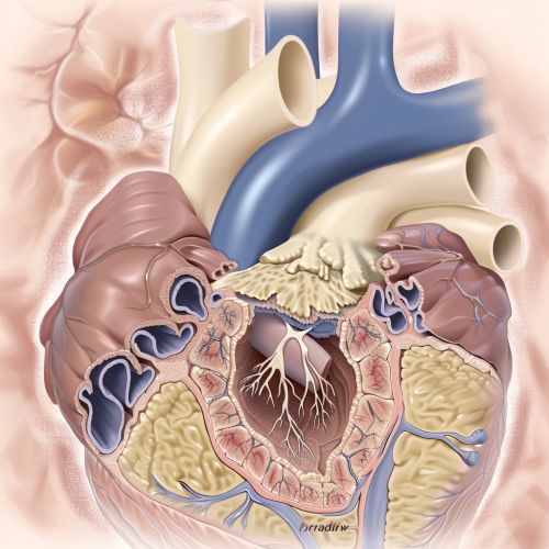
[[Image:Detail-97397.jpg|thumb|center|A medical illustration showing the anatomy of the heart with a focus on the narrowed pulmonary valve.|class=only_on_mobile]] | |||
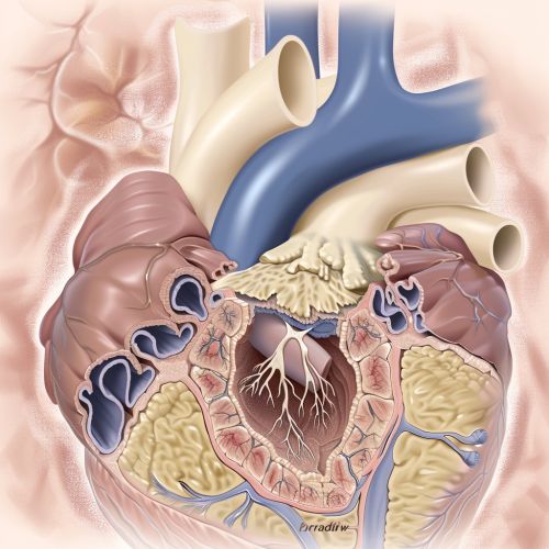
[[Image:Detail-97398.jpg|thumb|center|A medical illustration showing the anatomy of the heart with a focus on the narrowed pulmonary valve.|class=only_on_desktop]] | |||
== Prognosis == | == Prognosis == | ||
Latest revision as of 10:50, 29 July 2024
Overview
Pulmonary valve stenosis (PVS) is a congenital or acquired condition characterized by the narrowing of the pulmonary valve, which impedes blood flow from the right ventricle to the pulmonary artery. This condition can range from mild to severe and may be associated with other congenital heart defects. PVS can lead to increased right ventricular pressure, right ventricular hypertrophy, and potentially right heart failure if left untreated.
Etiology
Pulmonary valve stenosis is most commonly congenital, arising from abnormal development of the pulmonary valve during fetal growth. The exact cause of this abnormal development is often unknown, but it can be associated with genetic syndromes such as Noonan syndrome. Acquired causes of PVS are rare but can include rheumatic fever, carcinoid syndrome, and infective endocarditis.
Pathophysiology
The pathophysiology of PVS involves the obstruction of blood flow from the right ventricle to the pulmonary artery due to the narrowed valve. This obstruction increases the pressure within the right ventricle, leading to right ventricular hypertrophy as the heart works harder to pump blood through the stenotic valve. Over time, this increased workload can result in right ventricular dysfunction and heart failure.
Clinical Presentation
The clinical presentation of PVS varies depending on the severity of the stenosis. Mild cases may be asymptomatic and discovered incidentally during routine examinations. Moderate to severe cases may present with symptoms such as: - Dyspnea (shortness of breath) - Fatigue - Chest pain - Syncope (fainting) - Cyanosis (bluish discoloration of the skin)
Physical examination may reveal a systolic ejection murmur, best heard at the left upper sternal border, and a palpable thrill.
Diagnosis
The diagnosis of PVS typically involves a combination of clinical evaluation, imaging studies, and sometimes invasive procedures. Key diagnostic tools include: - **Echocardiography**: This is the primary imaging modality used to assess the structure and function of the pulmonary valve and right ventricle. Doppler echocardiography can measure the pressure gradient across the valve. - **Electrocardiography (ECG)**: May show signs of right ventricular hypertrophy or right atrial enlargement. - **Chest X-ray**: Can reveal right ventricular enlargement and post-stenotic dilation of the pulmonary artery. - **Cardiac catheterization**: Used in certain cases to directly measure the pressure gradient across the pulmonary valve and to assess the severity of the stenosis.
Management
The management of PVS depends on the severity of the condition and the presence of symptoms. Treatment options include:
Medical Management
Mild cases of PVS may not require any specific treatment and can be managed with regular monitoring. In symptomatic or severe cases, medical management may include: - **Beta-blockers**: To reduce heart rate and myocardial oxygen demand. - **Diuretics**: To manage symptoms of heart failure.
Interventional and Surgical Management
- **Balloon Valvuloplasty**: This is the first-line treatment for moderate to severe PVS. It involves the insertion of a balloon catheter through the femoral vein to the pulmonary valve, where the balloon is inflated to widen the valve opening. - **Surgical Valvotomy**: Indicated in cases where balloon valvuloplasty is not feasible or unsuccessful. It involves surgical incision of the valve to relieve the obstruction. - **Pulmonary Valve Replacement**: Reserved for cases with severe valve damage or when other interventions are not effective. This can be done using a bioprosthetic or mechanical valve.


Prognosis
The prognosis for individuals with PVS depends on the severity of the stenosis and the success of treatment. Mild cases generally have an excellent prognosis with normal life expectancy. Moderate to severe cases, if treated effectively, also have a good prognosis, although there may be a need for ongoing monitoring and potential re-intervention.
Complications
Potential complications of untreated or severe PVS include: - Right ventricular hypertrophy and failure - Arrhythmias - Infective endocarditis - Sudden cardiac death
Epidemiology
PVS is a relatively rare congenital heart defect, accounting for approximately 8-12% of all congenital heart disease cases. It occurs equally in males and females and can be associated with other congenital anomalies.
See Also
- Congenital Heart Disease - Noonan Syndrome - Balloon Valvuloplasty - Right Ventricular Hypertrophy - Heart Failure
References
[Include references if available]
