MUGA scan: Difference between revisions
(Created page with "== Overview == A **MUGA scan** (Multiple Gated Acquisition scan) is a type of nuclear medicine imaging test that evaluates the functioning of the heart's ventricles. This diagnostic tool is particularly useful for assessing the heart's pumping efficiency and is often employed in the management of patients with heart disease, especially those undergoing chemotherapy, which can affect cardiac function. == History and Development == The development of the MUGA scan dates...") |
No edit summary |
||
| Line 49: | Line 49: | ||
The patient lies on a table under the gamma camera, and ECG leads are attached to monitor the cardiac cycle. The gamma camera then captures images of the heart at various phases of the cardiac cycle. The data is processed to generate images and calculate functional parameters. | The patient lies on a table under the gamma camera, and ECG leads are attached to monitor the cardiac cycle. The gamma camera then captures images of the heart at various phases of the cardiac cycle. The data is processed to generate images and calculate functional parameters. | ||
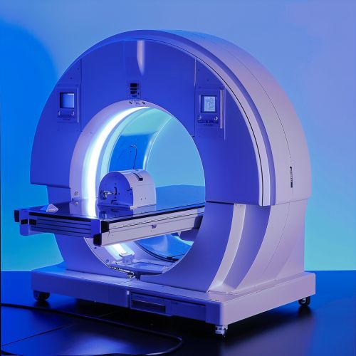
[[Image:Detail-93029.jpg|thumb|center|Gamma camera used in nuclear medicine imaging.]] | |||
== Interpretation of Results == | == Interpretation of Results == | ||
Revision as of 21:39, 21 June 2024
Overview
A **MUGA scan** (Multiple Gated Acquisition scan) is a type of nuclear medicine imaging test that evaluates the functioning of the heart's ventricles. This diagnostic tool is particularly useful for assessing the heart's pumping efficiency and is often employed in the management of patients with heart disease, especially those undergoing chemotherapy, which can affect cardiac function.
History and Development
The development of the MUGA scan dates back to the 1970s when advancements in nuclear medicine allowed for more precise imaging techniques. The introduction of gated blood pool imaging provided a significant leap in cardiac diagnostics, enabling clinicians to evaluate ventricular function with greater accuracy. Over the decades, the technology has evolved, incorporating more sophisticated imaging software and hardware, which has enhanced its diagnostic capabilities.
Technical Aspects
Radiopharmaceuticals
A MUGA scan involves the injection of a radiopharmaceutical, typically technetium-99m labeled red blood cells, into the patient's bloodstream. The radiopharmaceutical emits gamma rays, which are detected by a gamma camera to create detailed images of the heart.
Imaging Process
The imaging process is synchronized with the patient's electrocardiogram (ECG) to capture images at specific points in the cardiac cycle. This synchronization, or "gating," allows for the assessment of the heart's ejection fraction, wall motion, and other functional parameters. The images are acquired over several cardiac cycles to ensure accuracy and reproducibility.
Equipment
The primary equipment used in a MUGA scan includes a gamma camera, an ECG machine, and a computer system for image processing. The gamma camera detects the gamma rays emitted by the radiopharmaceutical and converts them into electrical signals, which are then processed to form images.
Clinical Applications
Cardiac Function Assessment
One of the primary uses of a MUGA scan is to evaluate the ejection fraction (EF) of the heart, which is a measure of how much blood the left ventricle pumps out with each contraction. An EF below 50% may indicate heart failure or cardiomyopathy.
Chemotherapy Monitoring
MUGA scans are frequently used to monitor cardiac function in patients undergoing chemotherapy, particularly with drugs known to have cardiotoxic effects, such as anthracyclines. Regular monitoring helps in early detection of cardiac dysfunction, allowing for timely intervention.
Preoperative Evaluation
In patients scheduled for major surgery, a MUGA scan may be performed to assess cardiac function and determine the risk of perioperative cardiac complications. This is especially important in patients with known heart disease or those undergoing procedures that place significant stress on the heart.
Procedure
Preparation
Prior to a MUGA scan, patients are usually advised to avoid caffeine and certain medications that may interfere with the test. The procedure itself is non-invasive and typically takes about 1-2 hours.
Injection of Radiopharmaceutical
The first step involves the intravenous injection of the radiopharmaceutical. After the injection, there is a waiting period to allow the radiopharmaceutical to circulate and bind to the red blood cells.
Image Acquisition
The patient lies on a table under the gamma camera, and ECG leads are attached to monitor the cardiac cycle. The gamma camera then captures images of the heart at various phases of the cardiac cycle. The data is processed to generate images and calculate functional parameters.

Interpretation of Results
The results of a MUGA scan are interpreted by a nuclear medicine specialist or a cardiologist. Key parameters evaluated include:
- **Ejection Fraction (EF):** A measure of the percentage of blood ejected from the left ventricle with each heartbeat.
- **Wall Motion:** Assessment of the movement of the heart walls during contraction and relaxation.
- **End-Diastolic and End-Systolic Volumes:** Measurements of the volume of blood in the ventricles at the end of filling and contraction phases, respectively.
Abnormalities in these parameters can indicate various cardiac conditions, such as heart failure, cardiomyopathy, or ischemic heart disease.
Advantages and Limitations
Advantages
- **Non-Invasive:** The procedure does not require surgery or catheterization.
- **High Accuracy:** Provides precise measurements of cardiac function, particularly ejection fraction.
- **Reproducibility:** Results are highly reproducible, making it useful for serial monitoring.
Limitations
- **Radiation Exposure:** Involves exposure to a small amount of ionizing radiation.
- **Availability:** Requires specialized equipment and trained personnel, which may not be available in all healthcare settings.
- **Cost:** Can be expensive compared to other imaging modalities.
Alternatives
Several alternative imaging modalities can be used to assess cardiac function, including:
- **Echocardiography:** Uses ultrasound waves to create images of the heart. It is widely available and does not involve radiation.
- **Cardiac MRI:** Provides detailed images of the heart using magnetic resonance imaging. It is particularly useful for assessing myocardial tissue characteristics.
- **Cardiac CT:** Uses computed tomography to obtain detailed images of the heart and coronary arteries. It is often used for coronary artery disease assessment.
Future Directions
Advancements in nuclear medicine and imaging technology continue to enhance the capabilities of MUGA scans. Research is ongoing to develop new radiopharmaceuticals and imaging techniques that provide even greater accuracy and diagnostic value. Additionally, the integration of artificial intelligence and machine learning in image analysis holds promise for improving the interpretation and clinical utility of MUGA scans.
